
The respiratory muscles or breathing pump muscles form semi-rigid bellows around the lungs in the chest. It is a complex arrangement of all the muscles which are attached to the rib cage, and help in generating the breathing action. These muscles include inspiratory muscles which cause the thoracic cavity to expand or induce inhalation and expiratory muscles which compress the thoracic cavity or induce exhalation. The basic structure of these muscles of respiration is the same as the other skeletal muscles of the body and they work in sync to expand or compress the chest while breathing.
What Are the Muscles of Respiration?
1. Diaphragm
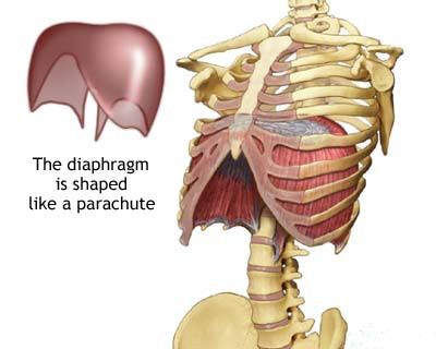 The major muscle responsible for helping us breathe is the diaphragm. It separates the thoracic and abdominal cavity by its thin and dome shaped structure. When the diaphragm contracts, its central portion moves downwards and sides move upwards and cause inhalation. The opposite action causes exhalation. This muscle is also responsible for helping in expelling vomit, feces and urine by applying pressure in the abdomen. It also prevents acid reflux by putting pressure on the esophagus.
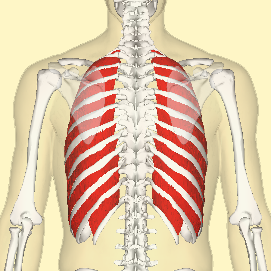 2. Intercostal Muscles
Intercostal muscles are one of the most important muscles of respiration and run along the diaphragm. They are attached between the ribs, and allows for changes in the width of the rib cage. Among the 3 layers of the intercostal muscles, the external ones are most important for respiration. They are placed in such a manner that when they contract, the ribs are raised and assist in inhalation.
3. Accessory Muscles of Respiration
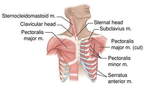 These muscles of respiration do not actively play a part in breathing. The sternocleidomastoid and the scalene muscles are considered as accessory muscles, and they help in elevating the rib cage. When a person is quiet breathing, the scalene muscles are active while sternocleidomastoid remains quiet. When respiration increases, the latter becomes active as well. Some other neck muscles are also considered as accessory muscles of respiration.
4. Muscles of Exhalation
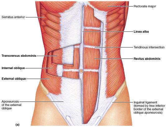 When a person is involved in quiet breathing, there is not much effort required by the muscles during exhalation. It is caused by the recoil of the thoracic wall. When there is forceful exhalation required, abdominal wall muscles contract. This can happen when the elasticity of the lungs reduces. These muscles reduce the volume of the thoracic cavity. Internal intercostal muscles also help in adding force to exhalation.
Here is a video explaining muscles involved in breathing process:
How Does the Breathing Process Develop?
To make it simple, breathing process is a process of taking in oxygen and expelling out carbon dioxide. When you inspire, air with rich oxygen runs through the mouth and nose, and then down to lungs via windpipe. Alveoli, the air sacs in the lungs, can transfer oxygen to the blood, while the bloodstreams is absorbing the oxygen and transferring the carbon dioxide to the air sacs. Therefore, when you expire, the carbon dioxide and other gaseous wastes are expelled out of your body. With the muscles of respiration, the respiration process happens in the following ways:
1. Inspiration
Inspiration or inhalation is an active process which requires contraction of the skeletal muscles. The diaphragm creates pressure difference between the abdominal cavity and the intra-pleural space, while there is some tension in the diaphragm. It is responsible for 2/3 of the inspired volume, during quiet breathing.
Intercostal muscles (particularly the external intercostal muscles) contract and cause the rib cage to rise. Thus, the dimension of the chest is increased in an anterior to posterior manner. That's how the inhalation process develops.
2. Expiration
Expiration or exhalation can be of two types – quiet breathing and forced expiration. Quiet breathing, in a passive pattern, happens due to the elastic recoil when the inspiration muscles relax. It is the normal breathing when at rest.
During forced expiration process, the internal intercostal muscles are in action, and lower the rib cage along with the abdominal muscles, which increase the abdominal pressure, as the diaphragm forces upwards.
3. Force Magnitude
Force magnitude has 2 aspects – quiet breathing and maximum respiratory effort. In quiet breathing, the inspiration muscles generate pressure which is equal to -5 cmH2O. While during expiration, the elastic forces act passively and generate 5 cmH2O, which decreases to 0 cmH2O when the chest comes back to its relaxed position.
In maximum respiratory effort, the inspiration pressure is -100 cm H2O, and the expiration pressure is 100 cm H2O. This interferes with cardiac output, especially venous return and requires extreme effort.
Valsalva maneuver is something which gives maximum expiratory effort, causing maximum expiratory pressure and happens with the glottis closed.
Here is a 3D visual illustration of how the muscles of respiration function in the breathing process.