
The synapse or connection between a motor neuron and a skeletal muscle is known as neuromuscular junction. Communication happens between the neuron and muscle via nerve cells. Due to this communication or transmission of signal, the muscle is able to contract or relax. It is the most widely studied synapse and it is comparatively easier to understand and analyze as well.
We shall look in detail about the junction, what it consists of, what are its functions as well as some disorders associated with the junction.
What Is Neuromuscular Junction?
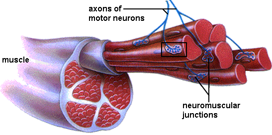
 A motor neuron is responsible for causing a skeletal muscle to contract, by stimulating it. The gap or space present between this motor neuron and the skeletal muscle cell is called as a synapse. This synapse, specifically between the skeletal muscle cell and motor neuron is called neuromuscular myoneural or junction. Myo means Muscle and Neural means Nerves. When an impulse travels between this space, muscle contraction happens.
There are around 100-500 trillion such connections in the human brain, between two nerves or nerves and glands. In this article we will discuss only the junction.
Structure of Neuromuscular Junction
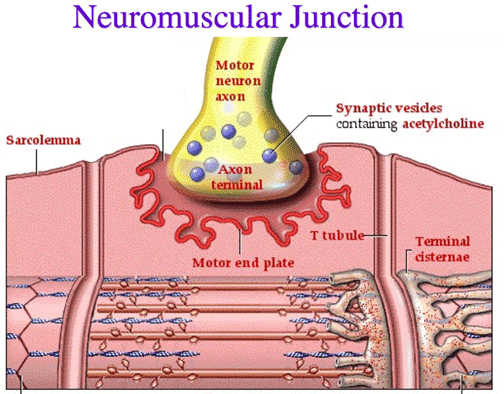
As we have read, the junction consists of a neuron and a skeletal muscle cell. The neuron in the combination is referred to as spinal motor neuron. The motor neurons, who originate from the spinal cord, innervate the skeletal muscle fibers. The innervation happens by very fine processes of the axon. The synapses are present along these processes and are also known as motor endplate, because of its specific structure.
The junction synapse has 3 characteristic features:
- There are two membranes called the pre and post synaptic membranes. There exists a distinct space between these membranes and is known as Synaptic Cleft.
- High density of small spherical vesicles are present, which contain neurotransmitter substances.
- A thickened post synaptic membrane is present, which has high density of receptors that are responsible for binding the chemical substances which transmit the signal from the presynaptic neuron.
Functions of Neuromuscular Junction
Before going in detail, let us remember the broad action of this junction. It is to act like a bridge between a neuron and muscle cell to transmit the signals.
Motor Neuron—Synaptic Cleft—Skeletal Muscle Cell
Calcium makes an entry in the excited motor neuron, which in turn causes exocytosis of the neurotransmitter. Which means the neurotransmitter gets transported to the only place available – the synaptic cleft. Acetylcholine is the neurotransmitter secreted by the somatic motor neurons.
There are receptors of acetylcholine present in the skeletal muscle cells. So this secreted acetylcholine then passes the cleft by diffusion and bind with the receptors. They are like puzzle pieces which fit or key which opens the door. This process opens an ion channel through which sodium ions pass to the muscle cells. At that time potassium ions diffuse out of muscle cells. However, the amount of Sodium ions entering is higher than the number of potassium ions leaving. Once the sodium ion reaches the muscle cell, it depolarizes, because it is a positive ion. This causes the skeletal muscle cell membrane to get excited and contract. This is referred to as Action Potential being triggered.
The acetylcholine does not remain in the synaptic cleft of the neuromuscular junction forever. This is to ensure that it does not cause over contraction of the muscle or keep the muscle contracted for longer than required. An enzyme called as Acetylcholinesterase is responsible in acting as a catalyst in breaking down the acetylcholine present in the synaptic cleft. It gives rise to acetate and choline, which are then transported back to the synaptic cleft, where they are re-synthesized. This is called as Active Reuptake.
For clearer demonstration of the junction, watch a video below:
Neuromuscular Junction Disorders
Some of the disorders affecting the junction are Myasthenia Gravis, Botulism, Eaton-Lambert syndrome etc. The junction can also malfunction when exposed to certain antibiotics, organophosphates which are type of insecticides, curare which is a toxin derived from plants and gases used in chemical warfare. Some of these things work by preventing the breakdown of acetylcholine after the transmission of the nerve impulse. While some of the disorders cause over activity of the muscles as described below:
- Stiff person syndrome: Antibodies attack nerve cells – responsible for muscle regulation - present in the brain and spinal cord and cause them to be continuously stimulated. Due to this they become stiff.
- Isaacs syndrome: In this the nerves keep sending impulses to the muscles which causes them to be over stimulated and ultimately stiffen. They also tend to twitch, due to which they find exercising and normal activities difficult to perform.