The human heart is composed of four chambers i.e. right and left atria and left and right ventricles. The right ventricle is responsible for pumping the oxygenated blood throughout the body to ensure the delivery of adequate oxygen in the tissues.
Bundle branch block refers to an obstruction or delay in the pathway of electrical impulse flowing to the right or left bottom side of ventricles or chambers in your heart, as a result, the pumping of heart is affected and conduction is slowed. On the basis of anatomical location, this bundle branch block may be of two major types: right bundle branch block and left bundle branch block.
What Is Left Bundle Branch Block?
LBBB, also called left bundle branch block, is a condition in which there is a conduction abnormality in heart. It is important to mention that this abnormal conduction can also be observed and seen in an ECG (electrocardiogram). The condition is characterized by delay in electrical impulses in left ventricle of heart due to which the left ventricle contracts later than the right ventricle.
Left bundle branch block is usually observed due to two significant reasons: an underlying cardiac disease and decreased efficiency of cardiac muscle to work and pump out the blood (it is present in patients who are already suffering from heart related disorders).
Causes and Symptoms of Left Bundle Branch Block
Causes
Some prominent causes of left bundle branch block include:
- Myocardial infarction
- CAD (coronary artery disease)
- Aortic stenosis
- Cardiomyopathy
- Lyme disease
- Aortic root dilatation and aortic root reconstruction
Symptoms
Most patients do not show any characteristic symptoms. In fact, in many cases the patient is not even aware of his condition that he is suffering from bundle branch block due to absence of any specific symptom. However in severe cases, following symptoms are reported:
- Syncope (fainting and dizziness)
- Presyncope (feeling of faintness but actually no faintness is present, the patient experience light headedness only)
If you have the above symptoms, you'd better visit your doctor to rule out any underlying serious issues. Or if you have been diagnosed with cardiac disease especially a conduction disorder, you should appoint regular follow-up medical visits with your doctor, which ensures that the doctor is familiar enough to your medical history in handling an emergency.
How Does Left Bundle Branch Block Affect Your Health?
1. Left Bundle Branch Block and Underlying Heart Disease
Almost 6% of old patients aging 80 are affected by the left bundle branch block. A study conducting on patients with left bundle branches block reveals that as the age of the patient progresses the incidence of developing cardiac diseases such as hypertension and cardiomegaly increases. Study also found that approximately 89% of the patients have developed multiple cardiac diseases and were diagnosed with heart failure, coronary artery disease, etc. However, in patients of lower age group the incidence of developing cardiac diseases is comparatively lower.
Patients with LBBB must be evaluated by a health care practitioner at regular intervals. Patients with high risk factors must maintain a healthy nutrition and lifestyle.
2. LBBB and the Efficiency of the Heartbeat
There is a perfect balance and coordination between the two ventricles of heart in normal heart conduction. In case of left bundle branch block, the left ventricle of heart receives later conduction and impulse signals than the right ventricle, which affects the coordination of heart muscles. As a result, the load on cardiac tissues to pump out blood increases.
A significant drop in cardiac efficiency is observed in patients of LBBB associated with other cardiac conditions such as heart failure, and the ejection fraction is observed to be lower than 35 percent.
How to Diagnose Left Bundle Branch Block

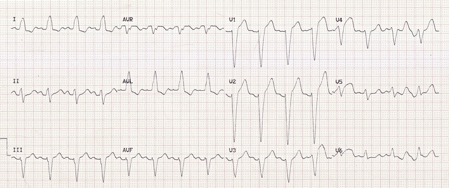
For diagnosis of LBBB, as the chart shows below, you will need to confirm the following criteria:
- Patient have the QRS duration > 120 ms.
- There should be a dominant S wave in V1.
- The R wave peak time is prolonged and greater than 60 ms in V5-V6—the left precordial leads.
- Q waves are absent in lateral leads—I, V5-V6. And small Q waves can also appear in aVL.
- R wave is broad and monophasic in lateral leads—I, aVL, V5-V6.
Some associated diagnostic features include:
- R wave progression is poor in chest leads.
- T waves and ST segments are observed in opposite directions to vector of QRS complex, this feature is termed as Appropriate Discordance.
- Left axis is deviated.
Left bundle branch block can be easily diagnosed through electrocardiogram, the QRS complex interval is observed to be greater than 120 milliseconds at lead V1.
If the QRS complex is deflected down side then it may indicate the presence of left bundle branch block; while in case of upward deflection, right side bundle branch block is diagnosed.
Sometimes in case of fast heartbeat, a rate dependent left bundle branch block is observed. When a rate dependent LBBB occurs at heartbeat > 100 beats/min, it becomes difficult to diagnose this condition and differentiate it between LBBB and ventricular tachycardia, both of which can cause wide QRS complex.
To clear the distinction between the two, the Brugada Criteria for ventricular tachycardia diagnosis can help. At the below ECG, we can see a normal sinus rhythm following with a rapid ventricular response-which is assumed as atrial fibrillation. Under a faster heartbeat rate, the QRS complex will change to the morphology of LBBB. And the QRS morphology complex will return to normal when ventricular rate slows down and the sinus rhythm restores.
For better understanding please refer to the video below:
Is Left Bundle Branch Block Serious?
According to different researches, left bundle branch block is less complicated than right bundle branch block and can be treated easily. If no other underlying heart disease is present, no significant treatment is required. However, in presence of other heart diseases along with characteristic symptoms of LBBB, pacemaker should be placed for controlling and coordinating the conduction of impulses.
